
Today, there are many methods for making micro-scale structures. Most of those that can make 3-dimensional structures, including 3D printing, are top-down in the sense that components of a structure are carefully located one by one at a target position using a very sophisticated machine. On the other hand, in a bottom-up method, components are self-assembled to form a complex structure. In this project, we pursue a possible break-through in bottom-up construction of micro-scale structures, using DNA and magnetic beads.

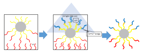
The goal of this project is to create “polychrome DNA beads”, whose sphere is separated into several regions, each covered with a different kind of ssDNA. “Polychrome DNA beads” are made from particles covered with the same ssDNA. Multiple kinds of ssDNAs with carboxyvinyl uracil (cvU) are prepared. If cvU is irradiated by UV light, it makes a covalent bond with an adjacent DNA molecule. Therefore, it should be possible to ligate a different kind of ssDNA with the strands on each region of the sphere. We then prepare DNA strands, called “DNA bridges”, which hybridize with those ligated DNA strands. By joining “polychrome DNA beads” by appropriate kinds of “DNA bridges”, we will be able to make accurate 3-dimensional structures in a bottom-up manner.
 There are two benefits in our approach compared with a similar prior study.*1
First, in our approach, any kind of material can be used for core of “polychrome DNA beads” if DNA strands can be attached on the surface of a particle as in avidin-biotin interaction. We can therefore make use of different features of materials for specific applications. We can also change the size of particles to make structures at different scales.
Second, only by changing the direction of UV light irradiation, it is possible to control the regions to which ssDNAs are attached. This means that those regions are not limited to two hemispheres. We can allow more colors and more sophisticated shapes to make more complex 3-dimensional structure.
- Prior research: Formation of 1D and 2D Gold Nanoparticle Arrays by Divalent DNA-Gold Nanoparticle Conjugates
Yuichi Ohya et al, 2012

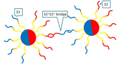
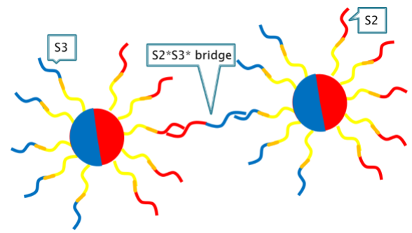
“Bi-chrome DNA beads” are the most basic “polychrome DNA beads” having two colors on two hemispheres. They are models for developing more general “polychrome DNA beads”.
Our “bi-chrome DNA beads” consist of magnetic beads, base DNA (named S1) and two kinds of DNAs (named S2 and S3). The “DNA bridge” consisting of S2* and S3* is used to join the beads, and a straight-line 1-dimentional structure is created.
We use magnetic beads for core of our “bi-chrome DNA beads”. This has some benefits. Magnetic beads can be localized at a specific area by applying a magnetic field to them. So we can increase the efficiency of self-assembly of “bi-chrome DNA beads”.

In the experiments, we use 3-micron magnetic beads and wells both coated with streptavidin. Three kinds of DNS2s are needed to make “bi-chrome DNA beads”, so we name them S1, S2 and S3, respectively.
Our method consists of three steps.
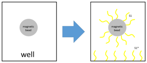
The first step is to attach S1 to beads and S1* to a well.
We attach S1 with biotin at 5’ end to the beads, and S1* to the well by avidin-biotin interaction.
Then we put the beads into the well and hybridize S1 and S1*.
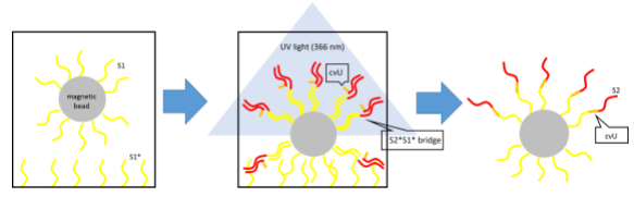
 The second step is to connect S2 with S1 on beads.
In a separate tube, we hybridized S2*S1* and S2 with cvU. S2 and S1 are not connected just as they are, so we use S2*S1* as a support.
Note that cvU, which stands for carboxyl vinyl uracil, is a photo reactive base, and is connected to an adjacent DNS2 when irradiated with the light whose wave length is 366nm.
S2 with cvU, which is hybridized with S2*S1*, is put in the well and hybridized with S1 on beads.
As a result, S2 with cvU and S2*S1* are hybridized with the copies of S1 on the beads that are not hybridized with the well.
We irradiate the well with the light whose wave length is 366nm. Then, cvU reacts to the light, and S1 on beads and S2 with cvU are ligated.
We irradiate the well for only a short time from the top of the well. So, only the upper hemisphere of the beads is exposed to the light. Then the copies of cvU only at the upper hemisphere of the beads react to light, and only DNA strands on the upper side are ligated. This is most important point in making “polychrome DNA beads” or “bi-chrome DNA beads”.
After that, by alkaline denaturation, S2*S1*s are removed from the beads, and the beads are removed from the well.
 The third step is to attach S3 to S1 on the beads.
We attach complementary S2 to another well and hybridize them with the processed beads. As in the second step, we hybridize S3 with S3*S1*, and add them to the well and hybridize them with S1. After irradiating the well with the light whose wave length is 366nm, we remove strands which are not ligated with S2 on the beads by alkaline denaturation. Then the construction of “bi-chrome DNA beads” is complete.
 We have not done an experiment for making multi-color DNA beads, but if the beads are immobilized to a well containing S2*, and irradiated with light from the side, we think it is possible to attach more kinds of DNA strands to the beads.

・Making “polychrome DNA beads”
We have succeeded in making “bi-color DNA beads”. The next step will be to make multicolor versions. We estimate that we can create up to hexa-colored beads. Using those beads, it would be possible to create even more complex 3-dimensional structures.
・Applying the characteristic features of magnetic beads
In our project, we used magnetic beads for core of our “bi-color DNA beads”. However, we could not make full use of characteristic features of magnetic beads. For example, magnetic beads generate heat by applying high-frequency magnetic waves, resulting in melting double strands of DNA. We may be able to use this feature for breaking once created structures.
Magnetic fields may also be useful for adjusting the density of “polychrome DNA beads”.
Both magnetic fields and high-frequency waves can operate by remote control and influence self-assembly of structures. That is, we may be able to control the process of self-assembly, resulting in different structures, or transforming self-assembled structures.